

Keratoconus and Corneal Irregularities
Keratoconus causes the cornea to become irregularly shaped, leading to blurred and distorted vision. Early detection and ongoing monitoring are essential for preserving vision and helping prevent disease progression.
Understanding Keratoconus
Keratoconus is a progressive eye condition that causes the cornea, the clear front surface of the eye, to gradually thin and bulge outward into a cone-like shape. As the cornea becomes more irregular, light entering the eye is distorted, leading to blurred vision, increased sensitivity to light, and difficulty achieving clear vision with glasses alone. Early detection and ongoing monitoring are important for preserving vision and managing disease progression.

Recognizing Keratoconus
Recognizing the early signs of keratoconus can help ensure earlier diagnosis and more effective management.
Blurred or distorted vision
Vision may appear blurry, shadowed, or warped, even with glasse
Increasing nearsightedness or astigmatism
Your prescription may become stronger more quickly than expected.
Frequent prescription changes
Glasses or contact lenses may need to be updated regularly.
Glare and halos around lights
Bright lights, headlights, and screens may appear to have rings or streaks.
Difficulty seeing at night
Night driving and low-light activities may become more challenging.
Light sensitivity
Eyes may become more sensitive to sunlight or bright indoor lighting.
Eye strain or headaches
Visual effort may lead to fatigue, discomfort, or frequent headaches.
Vision Not Fully Corrected by Glasses
Even with an updated prescription, vision may remain blurry or distorted.
Risk Factors for Keratoconus
Risk Factors for Keratoconus
Certain Genetic Conditions
Some genetic and chromosomal conditions have been associated with keratoconus. These conditions may affect corneal development or tissue strength, increasing the likelihood of corneal thinning and irregular shape changes.
Chronic Eye Rubbing
Frequent or vigorous eye rubbing places repeated stress on the cornea and is considered one of the most significant modifiable risk factors for keratoconus.
Asthma
Asthma is often linked with allergic conditions and may be associated with an increased risk of keratoconus, particularly when accompanied by chronic eye irritation or rubbing.
Severe Allergies or Eczema
Allergies and eczema can lead to chronic eye itching and rubbing, both of which have been associated with an increased risk of developing keratoconus.
Family History of Keratoconus
A family history of keratoconus increases the likelihood of developing the condition. Regular comprehensive eye exams can help detect early changes before noticeable vision problems occur.
Connective Tissue Disorders
Certain connective tissue disorders can affect the strength and structure of collagen throughout the body, including the cornea, which may increase susceptibility to keratoconus.
Corneal Imaging and Assessment
Advanced imaging is essential for detecting and managing keratoconus. These tests allow us to look closely at the shape and structure of the cornea, helping us identify early changes, make an accurate diagnosis, and monitor the condition over time.
Corneal topography to map the surface shape of the cornea and evaluate the full structure of the cornea in 3D
Optical coherence tomography (OCT) to assess corneal layers and thickness
Pachymetry and corneal thickness measurement
Wavefront analysis to detect visual distortions caused by irregular corneal shape
Understanding Your Keratoconus Evaluation
A comprehensive eye exam for keratoconus screens for progressive corneal thinning and irregular astigmatism to confirm diagnosis and monitor disease progression. The goal is to detect structural changes in the cornea, assess visual impact, and determine the appropriate management strategy.
Comprehensive Eye Examination
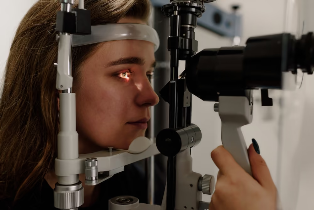
Your visit begins with a comprehensive eye exam to evaluate your vision, overall eye health, and any symptoms that may suggest keratoconus or other corneal irregularities. We also review your medical history, family history, and any previous changes in your glasses or contact lens prescription.
Corneal Mapping and Advanced Imaging
Advanced diagnostic technology, including corneal topography and tomography, is used to create a detailed map of the cornea. These measurements help detect subtle changes in corneal shape and identify keratoconus in its earliest stages.
Personalized Treatment Plan
After reviewing your results, we explain your findings and discuss the treatment options best suited to your needs. Depending on your condition, recommendations may include specialty contact lenses, monitoring, or co-management with a corneal specialist for treatments such as corneal cross-linking.
Ongoing Monitoring & Long-Term Care
Keratoconus requires regular follow-up to monitor changes in your vision and corneal health. Our team provides ongoing care, adjusts your treatment plan as needed, and works closely with specialists when appropriate to help preserve your vision over time.
Frequently Asked Questions (FAQs)
Find answers to common questions about keratoconus, specialty contact lenses, treatment options, and what to expect throughout your care.
Keratoconus is generally considered a progressive condition, meaning the cornea can continue to thin and change shape over time. The rate of progression varies significantly between individuals, some experience slow changes over many years, while others may notice more rapid progression. In some cases, the condition may stabilize later in life. Regular monitoring is essential to track changes and adjust treatment as needed.
There is currently no cure for Keratoconus. However, the condition can be effectively managed with early diagnosis and appropriate treatment. Advances in contact lens technology and procedures like corneal cross-linking have significantly improved the ability to stabilize the condition and enhance vision outcomes for many patients.
Keratoconus treatment depends on the severity and stage of the condition. In early stages, vision may be corrected with glasses or soft contact lenses. As keratoconus progresses, specialty contact lenses such as scleral lenses or rigid gas permeable lenses are often used to improve visual clarity. Corneal cross-linking is a treatment option designed to strengthen the cornea and help slow or stop the progression of the disease. In advanced cases, additional surgical options may be considered.
Frequent or vigorous eye rubbing is associated with an increased risk of keratoconus progression. Patients who have keratoconus or are at higher risk are encouraged to avoid rubbing their eyes, as repeated pressure on the cornea may contribute to further weakening and changes in corneal shape.
Monitoring frequency depends on the severity and progression of keratoconus. Most patients are seen every 6–12 months, with more frequent visits if the condition is progressing or after treatments like corneal cross-linking. We create a personalized follow-up plan to ensure appropriate ongoing care.
Many patients with keratoconus are able to wear specialty contact lenses, such as scleral or rigid gas permeable (RGP) lenses, which are custom-designed to provide clearer, more comfortable vision. During your evaluation, our providers will discuss your condition, explain the different lens options available, and recommend the solution that's best suited to your eyes, lifestyle, and visual needs.
Protect Your Vision with Early Detection
Schedule a comprehensive corneal evaluation to determine whether keratoconus or another corneal condition is affecting your vision. We'll provide a personalized care plan designed around your unique needs.