

Seeing the Whole Picture:
Ultra-Widefield Retinal Imaging
Protecting your vision starts with seeing the full picture. Advanced retinal imaging allows for earlier detection, more informed care, and a proactive approach to lifelong eye health.
Understanding the Retina
The retina is a thin, light-sensitive layer of tissue at the back of the eye that converts light into signals sent to the brain, making vision possible. Because the retina plays such a vital role in sight, diseases affecting this tissue can lead to permanent vision loss if not detected and treated early.
Many serious eye conditions, including diabetic retinopathy, macular degeneration, and others, can develop without noticeable symptoms in their early stages. Advanced retinal imaging helps identify subtle changes before vision is affected, allowing for earlier intervention and better long-term eye health outcomes.
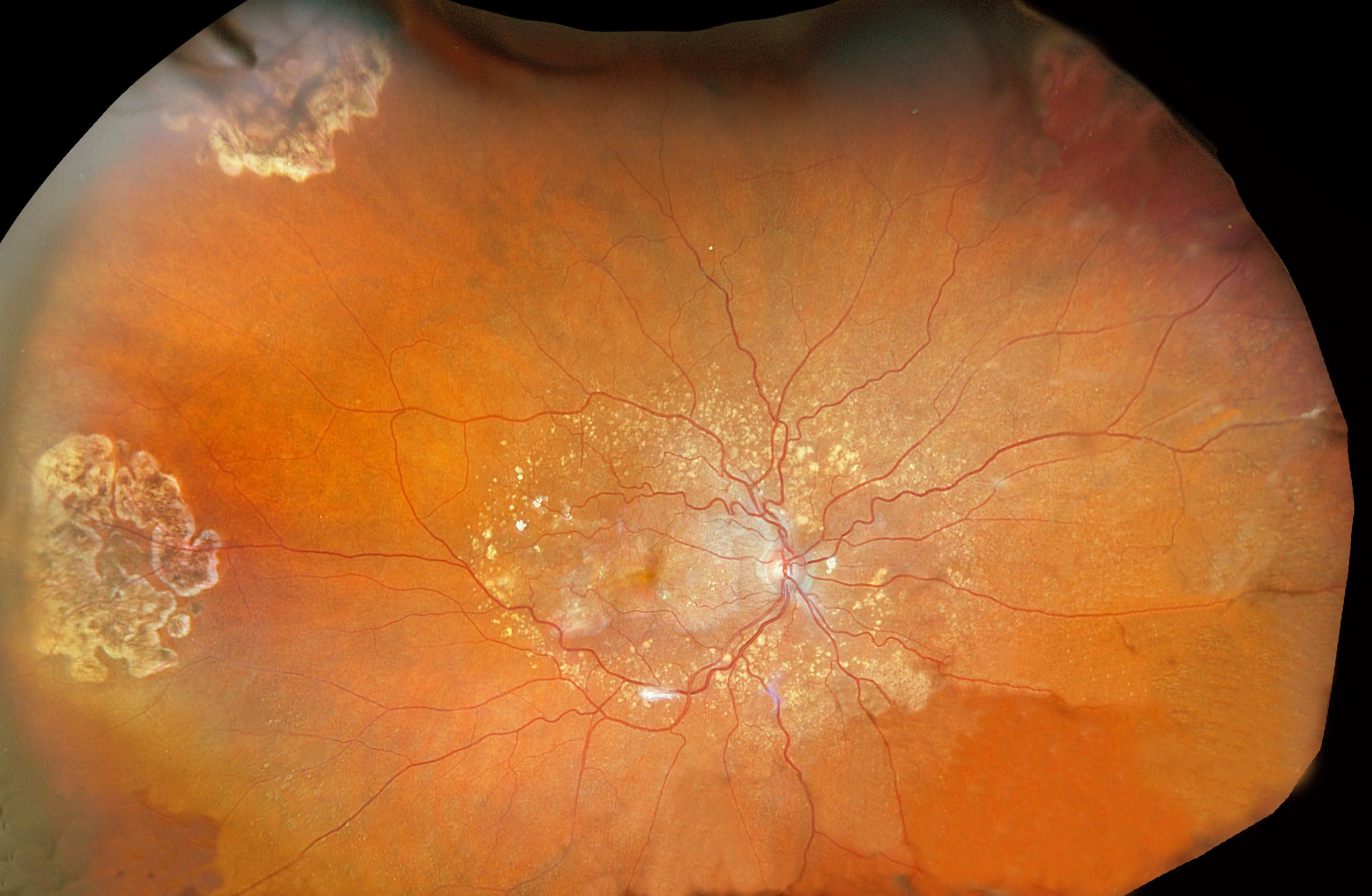
Advanced Retinal Imaging for Early Detection
Retinal imaging provides a comprehensive, high-resolution view of the eye’s most critical structures, including the retina, optic nerve, macula, retinal blood vessels, and peripheral retina. This advanced level of visualization allows for a more detailed assessment of ocular health than traditional examination methods alone.
By evaluating these structures with precision imaging technology, the doctor can identify subtle changes at the earliest stages and monitor for conditions that may require closer observation or timely intervention, supporting a more proactive and personalized approach to long-term eye health.
Comprehensive retinal evaluation
Permanent digital record
Early disease detection
Long-term health monitoring
Benefits of Optomap Retinal Imaging
Ultra-wide Field Imaging
Captures up to 200 degrees of the retina in a single image, providing a broader view of retinal health than traditional methods.
Digital Baseline Images
Compare images year to year and watch for subtle changes that signal disease progression.
Reduced Need for Dilation
Most patients can benefit from a comprehensive retinal evaluation without routine dilation, allowing them to return to daily activities without prolonged blurred vision or light sensitivity.
Quick and Comfortable
A detailed image is captured in seconds, allowing most patients to continue their day without the blurred vision
Early Disease Detection
Detects macular degeneration, diabetic retinopathy, hypertensive damage, retinal tears, and detachments before symptoms develop.
Detects Systemic Disease
Detects early signs of systemic diseases such as diabetes, high blood pressure, stroke, Alzheimer's and also certain cancers.
Diseases Detectable Through Retinal Imaging
Optomap imaging allows us to detect early signs of a wide range of eye diseases and systemic health conditions.

Age-Related Macular Degeneration (AMD)
Early changes in the macula that can affect central vision and progress over time.
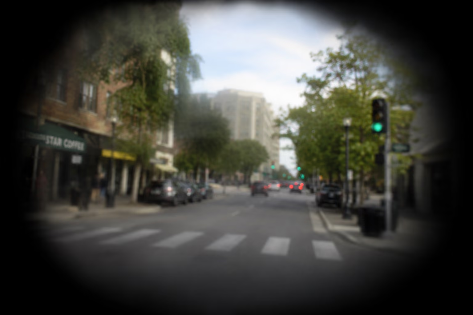
Hypertensive Retinopathy
Changes in retinal blood vessels related to high blood pressure.

Diabetic Retinopathy
Changes in retinal blood vessels caused by diabetes, including leakage, bleeding, or early vascular damage.
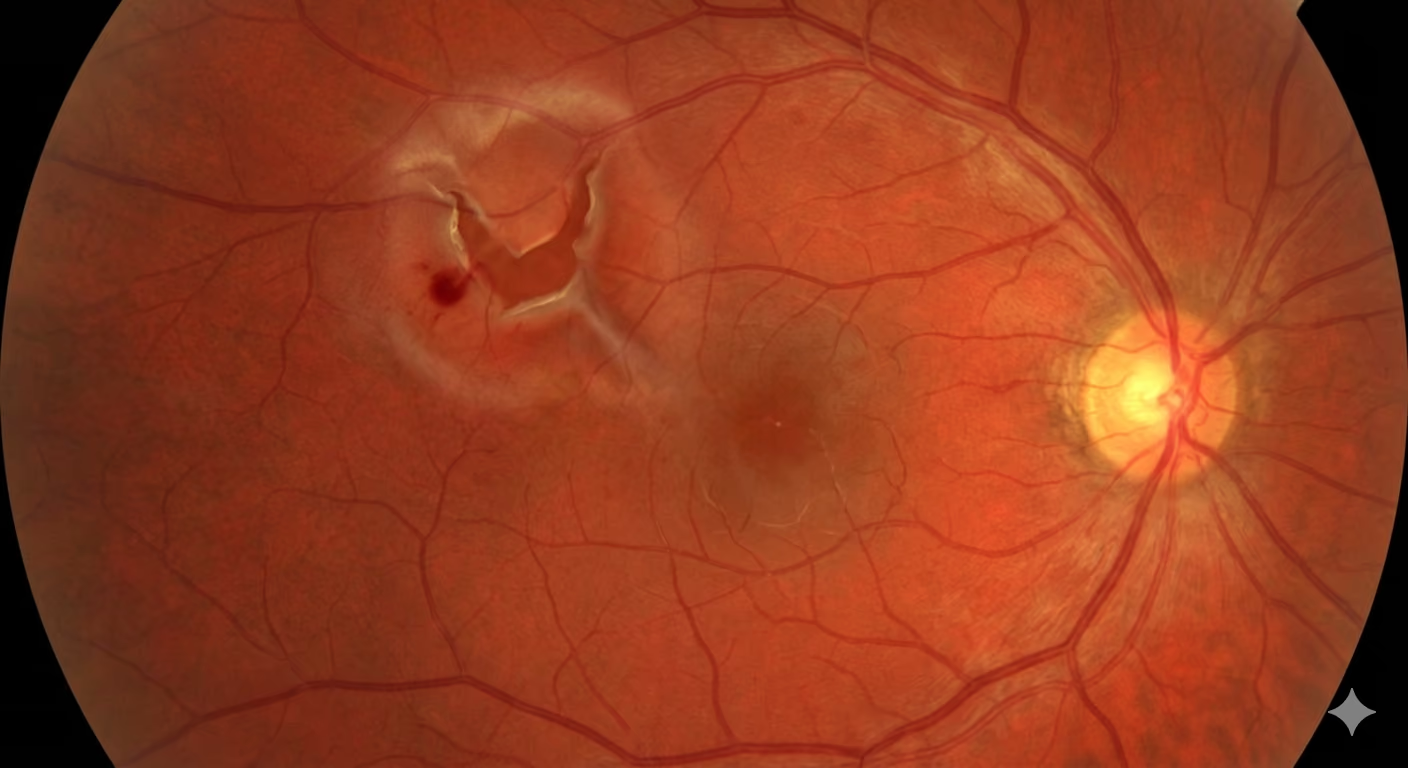
Retinal Tears
& Detachments
Small breaks or areas of weakness in the peripheral retina that may lead to vision-threatening detachment if untreated.
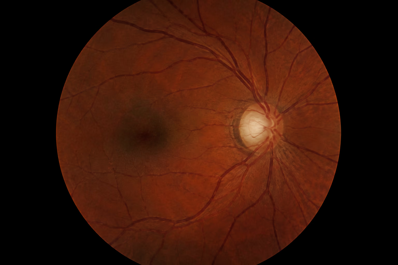
Glaucoma-Related Changes
Optic nerve appearance and structural changes that may indicate glaucoma or risk of progression.

Central serous retinopathy
Fluid builds up under the retina, leading to temporary blurry or distorted spot in central vision
What to Expect During Your Retinal Imaging
Ultra-widefield retinal imaging is a critical part of a comprehensive eye exam, allowing your doctor to evaluate the health of the retina in greater detail than traditional methods alone. The following steps outline how this advanced technology helps detect early changes and support proactive eye care.
Arrival and check-in
You’ll complete a brief health history and share any vision concerns related to both eye health and systemic health. Our team will answer questions and prepare you for your exam.
Ultra-widefield retinal imaging
Quick, non-invasive scan captures a detailed image of the retina, optic nerve, blood vessels, and peripheral retina in just seconds.
Doctor consultation and analysis
Images are reviewed for signs of retinal disease, glaucoma, diabetic eye disease, macular degeneration, and other changes that may affect vision and eye health.
Retinal images are reviewed with patients, providing a clear visual understanding of overall eye health and any areas of concern.
Plan and follow-up
Images are stored and compared over time, allowing subtle changes in retinal health to be detected and monitored annually. Based on findings, recommendations for further monitoring, testing, treatment, or a dilated eye exam may be discussed.
Does Optomap Replace Dilation?
Optomap retinal imaging provides a detailed view of much of the retina and is an excellent tool for documenting and monitoring retinal health. However, it does not completely replace a dilated eye examination.
Certain retinal conditions, peripheral retinal abnormalities, and specific medical situations may still require dilation to allow for a more thorough evaluation of the retina.
Depending on a patient's symptoms, medical history, risk factors, and clinical findings, one or both may be recommended to provide the most comprehensive assessment of eye health.

Frequently Asked Questions (FAQs)
Get answers about what to expect during with Retinal Imaging
Most patients benefit from annual retinal imaging as part of their routine comprehensive eye exam. Patients with diabetes, glaucoma, macular degeneration, high blood pressure, or a family history of eye disease may require more frequent imaging to monitor changes and protect long-term vision.
Ultra-widefield retinal imaging captures a much larger, more detailed view of the retina than a traditional exam. This allows your doctor to see areas that may be difficult to evaluate otherwise and identify early changes more effectively.
Retinal imaging can often provide detailed views of the retina without the need for dilating eye drops, making the exam more convenient for many patients. However, retinal imaging does not completely replace dilation in every situation. If your doctor needs a wider view of the retina or identifies an area that requires further evaluation, dilation may still be recommended. At Silo EyeCare, we use advanced retinal imaging technology to reduce unnecessary dilation whenever possible while still providing a thorough examination.
Yes. Digital retinal imaging is a safe, non-invasive procedure that uses light to capture detailed images of your retina. The test does not involve radiation, injections, or direct contact with the eye. Retinal imaging is appropriate for adults, children, and seniors and is commonly performed as part of a comprehensive eye exam.
Insurance coverage for retinal imaging varies depending on your medical history, diagnosis, and individual insurance plan. Some medical conditions may qualify for coverage, while other cases may involve an out-of-pocket fee. Our team will gladly discuss any costs and answer questions about your insurance benefits before your examination.
Yes. Retinal imaging is a quick and comfortable way to evaluate children's eye health. The images help document the health of the retina and optic nerve and establish a baseline for future examinations. Because the test is non-invasive and requires only a few seconds, many children tolerate retinal imaging very well.
See More. Know More.
Your eyes tell a story that goes beyond your vision alone. Advanced retinal imaging helps us detect, monitor, and protect your long-term eye health.